Macrophage Phenotype Identification Service
Overview Our Service Related Products Service Features Workflow Publications Scientific Resources Q & A

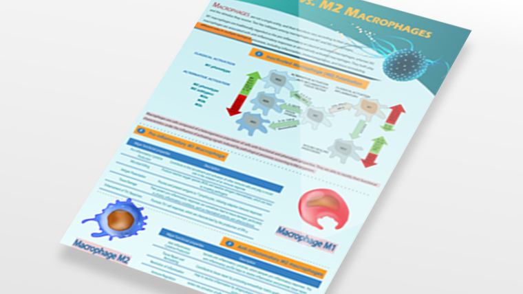
Macrophages, which accumulate in tissues during inflammation, may be polarized toward pro-inflammatory (M1), important effector cells, or tissue reparative (M2) phenotypes, capable of suppressing the function of M1 macrophages and influencing immunoregulation
and tissue repair. Identification of M1 and M2 macrophages would be beneficial for research, diagnosis, and monitoring of the effects of trial therapeutics in such diseases. Experienced in macrophage development and with a dedicated commitment
to the scientific community, Creative Biolabs has perfected our technical pipelines in the identification of M1 and M2 macrophages. We are happy to share our knowledge and passion in this field to facilitate our clients' research and project development.
Overview of Macrophage Phenotyping
Macrophages are the cornerstone cells of the immune system and play a key role in immunity, tissue homeostasis and pathology. Their phenotypic analysis provides important insights into the complexities
of immune regulation, disease progression and therapeutic response.
|
Phenotypes
|
Descriptions
|
Importance
|
|
M1
|
M1 macrophages are induced by pro-inflammatory signals such as IFN-γ and LPS and are associated with microbicidal activity and pro-inflammatory cytokine production.
|
-
Disease progression: Recognizing phenotypic changes can predict the progression of diseases such as cancer, cardiovascular disease, or chronic inflammation.
-
Therapeutic efficacy: Macrophage-targeted therapies, such as TAM reprogramming agents or M1/M2 modulators, rely on precise phenotypic understanding.
-
Immunomodulation: Understanding macrophage heterogeneity can improve immunotherapy outcomes, particularly in diseases where macrophages are dysfunctional.
|
|
M2
|
M2 macrophages are produced in response to anti-inflammatory stimuli such as IL-4 and IL-13. They are associated with wound healing, fibrosis, and immunosuppression.
Subtypes of M2 macrophages include:
-
M2a: promote tissue repair and antiparasitic responses
-
M2b: regulates inflammation
-
M2c: promote immunomodulation and stromal deposition
|
|
Other Specialized Phenotypes
|
-
Metabolically activated macrophages (MMe): Driven by high glucose and insulin levels, relevant in metabolic disorders.
-
Tumor-associated macrophages (TAMs): Exhibit a spectrum of phenotypes, often skewed towards immunosuppression and tumor promotion.
-
Resolution-phase macrophages: Facilitate inflammation resolution and tissue repair during the late phases of immune response.
|
Macrophage Polarization Phenotype Identification Service at Creative Biolabs
Creative Biolabs has established a cutting-edge Platform for macrophage development. Our scientists have accumulated extensive experience in applying real-time PCR, liquid chromatography-tandem mass spectrometry
(LC-MS/MS), western blot (WB), immunohistochemistry (IHC), and flow cytometry (FC) to identify classically activated M1 macrophages and alternatively activated M2 macrophages. A full range of macrophage markers, including multiple cell surface
markers, transcription factors, and cytokine profiles, are available for the identification of M1 and M2 macrophages.
For the identification of human M1 macrophages, IFN-γ, IL-1, IL-6, IL-10, IL-12, IL-23, TNF-α, CD16, CD32, CD64, CD68, CD80, CD86, CD369, inducible nitric oxide synthase (iNOS), signal transducer and activator of transcription 1 (STAT1), interferon regulatory
factor 5 (IRF5), Mer tyrosine kinase (MerTK), class II major histocompatibility complex molecules (MHC class II), and CXCL9 are reliable markers, while for mouse M1 macrophages, IFN-γ, IL-1, IL-6, IL-10, IL-12, IL-23, TNF-α, CD14, CD16, CD32,
CD64, CD68, CD80, CD86, CD204, CD369, IRF5, MerTK, MHC II, and Ly-6C are often used.

For the identification of human M2 macrophages, indoleamine 2,3-dioxygenase (IDO), IL-10, TGF-β, CD115, CD204, CD163, CD206, CD209, Fc epsilon Receptor I (FceR1), V-set and immunoglobulin domain-containing protein-4 (VSIG4), interferon regulatory factor
4 (IRF4), and signal transducer and activator of transcription 6 (STAT6) are reliable markers, while for mouse M2 macrophages, arginase 1 (Arg1), IDO, IL-10, TGF-β, CD14, CD115, CD163, CD204, CD206, CD209, colony-stimulating factor 1 receptor
(CSF1R), FceR1, YM1, IRF4, resistin-like molecule-alpha (RELM-α) and STAT6 are usually used.

As a long-term expert in the field of macrophage development, Creative Biolabs has rich expertise, which has been accumulated through our over a decade of experience. Integrate with other advanced platforms and technology,
we offer the most comprehensive analysis of M1 and M2 macrophages. If you are interested in our macrophage identification service, please feel free to contact us and further discuss it with our scientists.
Related Products
With deep expertise and a state-of-the-art technology platform, Creative Biolabs' R&D science team has designed and developed a range of useful products to assist our customers in macrophage phenotyping and other macrophage-related studies.
We offer different polarized macrophage subpopulations. In addition, our comprehensive product portfolio includes reagents for macrophage culture, M1/M2 macrophage differentiation,
and other related products that help our customers with their research.
|
Cat.No
|
Product Name
|
Product Type
|
|
MTS-0922-JF10
|
Human Macrophages, Alveolar
|
Human Macrophages
|
|
MTS-0922-JF99
|
Human M0 Macrophages, 1.5 x 10^6
|
Human M0 Macrophages
|
|
MTS-0922-JF52
|
C57/129 Mouse Macrophages, Bone Marrow
|
C57/129 Mouse Macrophages
|
|
MTS-0922-JF7
|
Human M2 Macrophages, Peripheral Blood, 10 x 10^6
|
Human M2 Macrophages
|
|
MTS-0922-JF34
|
CD1 Mouse Macrophages
|
CD1 Mouse Macrophages
|
|
MTS-1123-HM1
|
Macrophage Mannose Receptor 1 (MRC1) ELISA Kit, Colorimetric
|
ELISA Kit
|
|
MTS-1123-HM7
|
Macrophage Colony Stimulating Factor (MCSF) ELISA Kit, Fluorometric
|
ELISA Kit
|
|
MTS-1123-HM11
|
Monocyte To Macrophage Differentiation Associated Protein (MMA) ELISA Kit, Colorimetric
|
ELISA Kit
|
|
MTS-1123-HM9
|
Macrophage Expressed Gene 1 (MPEG1) ELISA Kit, Colorimetric
|
ELISA Kit
|
|
MTS-1123-HM15
|
Macrophage Chemokine Ligand 19 (CCL19) ELISA Kit, qPCR
|
ELISA Kit
|
|
MTS-1123-HM50
|
Macrophage Migration Inhibitory Factor (MIF) Competition ELISA Kit
|
ELISA Kit
|
Service Features
Comprehensive Phenotyping Panels
We utilize a broad range of molecular and cellular markers to differentiate macrophage subsets and characterize their activation states. Key markers and molecules assessed:
-
Surface markers
-
Cytokine profiles
-
Transcription factors
-
Metabolic indicators
Advanced Analytical Technologies
Our service employs cutting-edge tools to ensure precise and reproducible results.
-
Flow cytometry
-
RNA sequencing (RNA-seq)
-
Proteomics
-
Single-cell analysis
-
Immunohistochemistry (IHC)
Tailored Experimental Design
We recognize that research objectives vary widely. Our services include:
-
Customizable marker panels
-
Flexible sample types
-
Targeted data analysis
High-Quality Data Reporting
Our comprehensive data reports include:
-
Quantitative and qualitative analysis of macrophage subsets
-
Visual data presentations (e.g., heatmaps, clustering diagrams)
-
Detailed experimental methodology and result interpretation
Workflow of Macrophage Phenotype ldentification
Sample Preparation Phenotyping Panel Design Experimental Analysis Data Acquisition Data Interpretation Result Delivery
Publications
Rapid classification of macrophage phenotypes is challenging. David Dannhauser et al. investigated a method to classify polarized macrophage subtypes based on morphology. They compared
different machine learning algorithms to classify different macrophage phenotypes based on optical features obtained from a specially developed wide-angle static light scattering device. The main result was that they were able to identify
unpolarized macrophages from both M1 and M2 polarized phenotypes and distinguish them from naïve monocytes with an average accuracy of more than 85%.
In that study, they focused on intracellular differences in size and optical angle of macrophages. The optical cellular features obtained by their broad static light scattering method can significantly improve macrophage phenotypic studies by providing
a morphological characterization of the suspended cells.
 Fig. 1 Fluorescence and bright-field investigation of monocytes and M0, M1 and M2 macrophage phenotypes recovered from a healthy donor.1
Scientific Resources
Q & A
Q: What types of samples can I submit for analysis, and how should they be prepared?
A: We accept various sample types including isolated primary macrophages, cell culture samples, tissue sections, and biological fluids.
For cell suspensions, we recommend providing at least 1x10^6 viable cells in an appropriate transport medium. Tissue samples should be fresh frozen or properly fixed. We provide detailed sample preparation guidelines, including recommended
preservation protocols and shipping conditions, to ensure optimal sample quality upon arrival at our facility.
Q: Do you offer an analysis of temporal changes in macrophage populations over multiple timepoints?
A: Yes, we specialize in longitudinal studies of macrophage phenotype dynamics. We can analyze samples from multiple
timepoints and provide a detailed trending analysis showing phenotype transitions and population shifts. Our reports include statistical analysis of temporal changes, graphical representations of population dynamics, and interpretation of
potential biological significance.
Q: Can you help with experimental design and provide consultation for macrophage-related studies?
A: Yes, our team of experienced scientists offers comprehensive consultation services. We can assist with experimental
design, including selection of appropriate controls, timepoints, and analysis parameters. We provide guidance on sample preparation, handling, and storage to optimize results.
Q: What techniques do you use for phenotype identification, and how reliable are your results?
A: We employ a multi-platform approach combining flow cytometry, qRT-PCR, and immunofluorescence microscopy. Our flow cytometry
uses advanced spectral analysis to minimize artifacts. Results are validated through biological replicates and quality controls. Each sample is analyzed independently by two technicians, and we include standardized controls to ensure consistency
across experiments.
Q: What is your pricing structure?
A: We can provide detailed quotes based on specific project requirements and volume.
Reference
-
Dannhauser, David, et al."Single cell classification of macrophage subtypes by label-free cell signatures and machine learning." Royal Society Open Science 9.9 (2022): 220270. Distributed under Open Access license CC BY 4.0, without modification.


 Fig. 1 Fluorescence and bright-field investigation of monocytes and M0, M1 and M2 macrophage phenotypes recovered from a healthy donor.1


