Phenotype Difference Analysis Service by Flow Cytometry
Overview Our Service Related Products Service Features Publications Scientific Resources Q & A
Depending on microenvironmental stimuli, macrophages polarize into distinct phenotypes such as the classically activated M1 and the alternatively activated M2 types, each playing opposing roles in immune regulation, tissue remodeling, and pathogenesis of various diseases.
Understanding these phenotypic differences is crucial for advancing translational research, especially in oncology, infectious diseases, autoimmune disorders, and regenerative medicine. Creative Biolabs provides a comprehensive macrophage phenotype difference analysis service by flow cytometry, enabling clients to precisely define and compare macrophage subsets.
Phenotype Difference Analysis Among Macrophages
Many pathological processes rely on diverse macrophage populations, which vary greatly in shape, metabolism, expressed markers, and activities. Considering this, precise identification, counting, and phenotypic characterization of macrophage populations and phenotypes are necessary for understanding their pathophysiological roles and modeling the immune response involving macrophages.
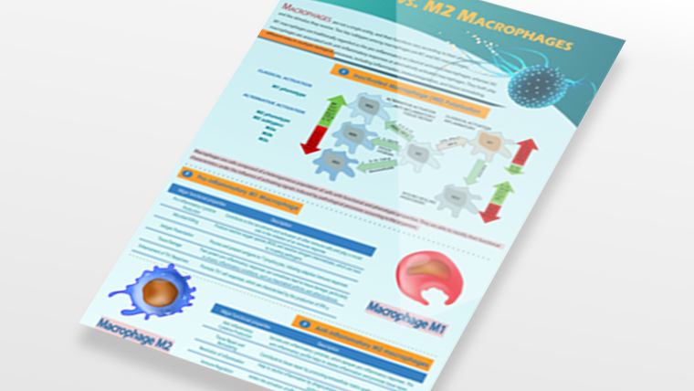
 Fig.1 The difference between M1 and M2 macrophages.1,3
Flow cytometry is an essential technique for immunophenotyping due to its ability to analyze multiple parameters simultaneously at the single-cell level. For macrophages, this technology facilitates:
-
High-throughput characterization of surface and intracellular markers
-
Quantitative analysis of heterogeneous cell populations
-
Identification of rare subsets and dynamic phenotype transitions
-
Functional assays involving cytokine profiling, phagocytic activity, and antigen presentation
Our Phenotype Difference Analysis Service by Flow Cytometry
Creative Biolabs has succeeded in providing a highly efficient and accurate phenotype difference analysis service by flow cytometry to serve as a useful tool to characterize macrophage phenotypes. We provide multiple types of cytometry to fulfill customers' diverse needs. At the same time, we also deliver a wide range of fluorochromes and several target molecules to design for your significant macrophage projects. In addition, we are confident in customizing the appropriate solutions if you have any other demands. At Creative Biolabs, we will try our best to deliver the best outcomes for every customer at a short turnaround.
As the core technology of our phenotype difference analysis service, we have already established a well-equipped flow cytometry platform, which is highly sensitive, high-throughput, and robust. The following are several types of well-developed cytometry that can be chosen for your projects:
-
Multicolor Conventional Flow Cytometry.
-
Spectral Flow Cytometry.
-
Mass Cytometry.
-
Multicolor Flow Cytometry.
Fluorochromes are the building blocks of signal detection for the flow cytometry technologies. Here we supply several fluorochromes to choose from for your distinctive demands:
-
Organic Small Molecules: fluorescein; cyanine dyes.
-
Natural Macromolecules: phycoerythrin (PE); allophycocyanin (APC); peridinin-chlorophyll-protein complex (PerCP).
-
Inorganic Quantum Dots (qDots).
-
Tunable Small Molecules: DyLight families.
-
Fluorescent Polymers.
Additionally, we also provide several well-identified marker molecules on macrophages for your convenience:
 Fig.2 Popular target molecules.
With Creative Biolabs' service, researchers gain access to robust, high-resolution tools for dissecting immune landscapes, identifying biomarkers, and accelerating the path from discovery to application.
Table 1 Core features of our service
|
Feature
|
Description
|
|
Marker Panel Customization
|
Tailored panels for M1/M2-specific markers
|
|
Multicolor Flow Cytometry
|
Up to 14-color panels for complex phenotype resolution
|
|
Intracellular Cytokine Staining
|
Optional add-on for functional phenotype classification
|
|
Species Flexibility
|
Human, mouse, rat, and other animal models supported
|
|
Cell Sorting Capabilities
|
Optional FACS sorting for downstream applications
|
|
Detailed Data Reporting
|
Quantitative plots, statistical summaries, and interpretation
|
Related Products
To gain insight into the function and status of macrophages, flow cytometry has become an indispensable analytical tool. Our analysis services are designed to provide researchers with high-precision cell characterization.
Additionally, our products such as macrophage products and cell culture media provide a superior growth environment for macrophage culture, promoting their physiological activity and function. By choosing our services and products, you'll receive a full range of efficient and reliable research support for your scientific endeavors.
|
Cat.No
|
Product Name
|
Product Type
|
|
MTS-0922-JF4
|
Human M2 Macrophages, Peripheral Blood (Age: 47), 2 x 10^6
|
Human M2 Macrophages
|
|
MTS-0922-JF99
|
Human M0 Macrophages, 1.5 x 10^6
|
Human M0 Macrophages
|
|
MTS-0922-JF5
|
Human M2 Macrophages, Peripheral Blood (Age: 21), 2 x 10^6
|
Human M2 Macrophages
|
|
MTS-0922-JF7
|
Human M2 Macrophages, Peripheral Blood, 10 x 10^6
|
Human M2 Macrophages
|
|
MTS-0922-JF34
|
CD1 Mouse Macrophages
|
CD1 Mouse Macrophages
|
|
MTS-0922-JF6
|
Human M1 Macrophages, Peripheral Blood (Age: 32), 5 x 10^6
|
Human M1 Macrophages
|
|
MTS-1022-JF1
|
B129 Mouse Bone Marrow Monocytes, 1 x 10^7 cells
|
Mouse Monocytes
|
|
MTS-0922-JF8
|
Human M1 Macrophages, Peripheral Blood (Age: 38), 5 x 10^6
|
Human M1 Macrophages
|
|
MTS-0922-JF9
|
Human M1 Macrophages, Peripheral Blood (Age: 30), 5 x 10^6
|
Human M1 Macrophages
|
Service Features

High-throughput Technologies
A single feature of multiple cells can be measured simultaneously. Up to 60 parameters can be routinely measured on a single cell.

High Precision
The intricacies of gene activity in individual cells can be unveiled. We strictly conduct quality control on the entire process to ensure high reliability and repeatability of experimental results.

Diversified Choices
The mysteries of gene expression at the single-cell level can be unlocked through various cutting-edge flow cytometry techniques and a wide range of fluorescent dyes.
Publications
To investigate the effects of exosomes released from adipose-derived mesenchymal stem cells (AdMSCs) on macrophages, the investigators isolated and cultured AdMSCs and human peripheral blood mononuclear cells (PBMCs) and assessed whether secreted exosomes could induce M2 polarization. Flow cytometry results revealed that AdMSCs increased the expression of M2 macrophage markers.
 Fig. 3 Expression of CD206 in PBMCs after coculture as assessed by flow cytometry.2,3
To confirm whether AdMSC induced the M2 phenotype in PBMCs, they assessed the expression rate of the M2 macrophage-specific marker CD206 using flow cytometry and immunofluorescence staining. Co-culture slightly increased CD206 expression in M2 macrophages.
Scientific Resources
Q & A
Q: What types of samples can I submit for macrophage phenotype analysis?
A: We accept a wide variety of sample types, including PBMCs, adherent macrophages from bone marrow or monocytes, and in vitro differentiated macrophages (e.g., M-CSF or GM-CSF derived). If needed, we can assist in macrophage enrichment or isolation using magnetic beads or FACS sorting prior to flow analysis.
Q: Can your service identify mixed phenotypes or transitional macrophage states?
A: Absolutely. One of the strengths of flow cytometry is its ability to detect cells expressing multiple markers simultaneously, revealing transitional or hybrid phenotypes. For example, cells co-expressing CD86 and CD206 may indicate a shift from M1 to M2 states. We also employ multivariate analyses (e.g., t-SNE, PCA) to visualize phenotype continuums and clusters, offering insights beyond binary M1/M2 classifications.
Q: Can I request customized panels or add cytokine detection in the same run?
A: Yes, we offer fully customizable multicolor panels tailored to your research questions. In addition to surface markers, we can incorporate:
-
Intracellular cytokines (e.g., TNF-α, IL-10, IL-12, TGF-β)
-
Phospho-protein detection (e.g., STAT signaling)
-
Live/dead staining, and viability profiling
-
Optional functional assays, such as phagocytosis markers (e.g., CD64)
Let us know your goals, and we will design the most efficient and cost-effective panel for your project.
Q: Is it possible to sort specific macrophage subsets for downstream applications?
A: Yes, we offer FACS-based cell sorting as an add-on. Sorted subsets can be returned to you in sterile condition for downstream applications such as:
References
-
Yao, Yongli, et al. "Macrophage polarization in physiological and pathological pregnancy." Frontiers in Immunology 10 (2019): 792. https://doi.org/10.3389/fimmu.2019.00792
-
Heo, June Seok, Youjeong Choi, and Hyun Ok Kim. "Adipose‐derived mesenchymal stem cells promote M2 macrophage phenotype through exosomes." Stem cells international 2019.1 (2019): 7921760. https://doi.org/10.1155/2019/7921760
-
Under Open Access license CC BY 4.0, without modification.


 Fig.1 The difference between M1 and M2 macrophages.1,3


 Fig. 3 Expression of CD206 in PBMCs after coculture as assessed by flow cytometry.2,3


